
A changing dark spot on skin should be medically assessed before it is treated as a cosmetic pigmentation problem. A change does not automatically mean cancer, but growth, a different shape or colour, bleeding, persistent itching or a lesion that does not heal can warrant attention. Do not try to remove it first and seek a diagnosis afterwards.
Start with an appropriate medical clinician, such as your GP or a dermatologist. They can decide whether examination, specialist referral or further investigation is needed. A general pigmentation treatment page or an online photograph comparison cannot establish that a changing lesion is safe to laser, peel or lighten.
Key takeaways
| Question | What to know |
|---|---|
| Does change prove cancer? | No. It is a reason for assessment, not a diagnosis. |
| Should cosmetic treatment come first? | No. Clarify what the lesion is before attempting removal or lightening. |
| What changes matter? | Growth, shape or colour change, new symptoms, bleeding or failure to heal. |
| Can a small spot still need review? | Yes. Size alone cannot rule out a concerning lesion. |
| What helps the appointment? | A timeline, previous photographs and any treatment history. |

Why should a changing spot be assessed?
Many skin lesions are benign, but a cosmetic label such as “sunspot” should not be assigned solely from colour. NHS guidance recommends medical review of moles that change or develop symptoms. A lesion can deserve attention even when it is not painful.
The important distinction is between noticing a change and diagnosing its cause. You do not need to decide whether it looks like a particular disease before booking. Describe what has changed and when, then allow the clinician to determine the next step.
What does the ABCDE guide mean?
AAD describes the ABCDE warning features used to help recognise possible melanoma. They are a prompt to seek assessment, not a home test that can confirm or exclude cancer.
| Question | What to know |
|---|---|
| A: asymmetry | One part has a different shape or appearance from the other. |
| B: border | The edge looks irregular or poorly defined. |
| C: colour | Colour varies within the lesion or has changed. |
| D: diameter | Larger size can be relevant, but concerning lesions may also be small. |
| E: evolving | The lesion changes or looks different from your other spots. |
You do not need all five features before asking for advice. A spot that behaves differently from your usual marks may be important even if it does not resemble a teaching photograph. Do not repeatedly delay an appointment while trying to satisfy a checklist.
Which symptoms should you report?
Describe bleeding, crusting, persistent itching, pain, a sore that does not heal or a lesion that heals and returns. Tell the clinician whether a symptom happened after accidental rubbing or appeared without an obvious injury. That detail is useful but does not independently establish the diagnosis.
If there is heavy bleeding that will not stop, significant infection symptoms or another acute concern, seek urgent medical care. Otherwise, arrange a timely medical appointment for a suspicious change rather than waiting for an aesthetic promotion or treating it as routine uneven skin tone.
Does skin tone change the need for assessment?
People of any skin tone can develop skin cancer. AAD guidance highlights that concerning lesions in darker skin can also occur on the palms, soles or around nails. A facial-pigmentation routine does not address every relevant site.
In a diverse Malaysian population, avoid using one photographic example as the only model of a warning sign. The lesion’s history, examination and individual circumstances matter. If a spot looks unusual for you, say so even when you cannot describe it using medical terminology.
Can acne marks or melasma be confused with other lesions?
A person can have ordinary post-inflammatory marks and a separate lesion at the same time. An old diagnosis for one area should not automatically be applied to every new spot. A changing isolated lesion may need a different assessment from a stable pattern left by previous acne.
Read our overview of facial pigmentation causes for general background, but do not use it to self-diagnose a changing mark. The purpose of educational content is to help you ask clearer questions, not to replace examination.
Why avoid laser, peels or removal before diagnosis?
An attempt to lighten or remove an undiagnosed lesion can alter its appearance and make the history more difficult to interpret. Do not use home acids, cautery tools, scraping or an unlabelled cream to “see whether it comes off”. A dark spot is not necessarily surface pigment that should be erased.
Tell any cosmetic practitioner that the lesion is changing and ask for medical assessment before treatment is considered. A recommendation to defer a procedure can be a useful safety decision. A familiar brand name or a previous successful cosmetic treatment does not establish that this lesion is suitable.
What might happen at the appointment?
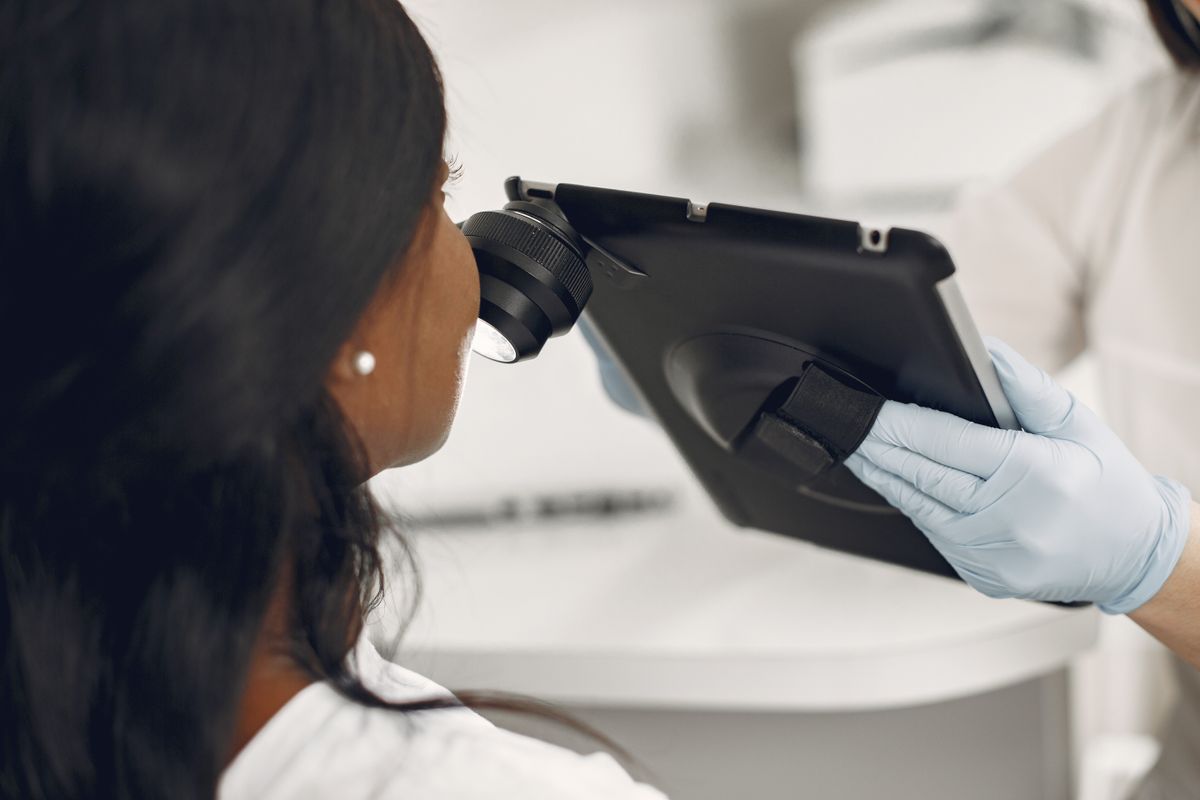
The clinician will take a history and examine the lesion. Depending on the findings and available expertise, they may discuss further examination, referral or biopsy. AAD explains that a skin biopsy and laboratory examination are used to establish a skin-cancer diagnosis. A referral or investigation does not by itself mean cancer has been confirmed.
Ask what the clinician thinks is likely, what remains uncertain and what the next step is intended to clarify. If observation is advised, ask for a defined follow-up plan and which changes should trigger earlier contact. Do not assume that “monitor it” means ignoring it indefinitely.
How can you prepare useful photographs?
Leeds Teaching Hospitals guidance discusses comparing lesions with baseline photographs and noting changes. Use similar lighting and include a scale if practical without pressing or injuring the skin. Keep the date so the clinician can understand the sequence.
Photographs support the history but cannot rule out a concerning lesion. Do not postpone assessment until you have a perfect image or several months of comparison. If the spot is already changing or symptomatic, arrange the appointment and bring whatever information is available.
What information should you bring?
- When the spot was first noticed and when the change began.
- Previous photographs, including ordinary images that incidentally show it.
- Symptoms such as bleeding, itching, pain or failure to heal.
- Any previous diagnosis, biopsy or treatment of the area.
- Medicines and relevant personal or family skin history.
- Products applied specifically to lighten or remove it.
- Your questions about examination, referral and follow-up.
If you are unsure whether it is new, say so. Memory is imperfect, and uncertainty is useful information. Avoid filling gaps with a guess that makes the history seem more definite than it is.

What if an online service says it is harmless?
A photograph-based opinion has limits. If the lesion continues to change, symptoms develop or the explanation does not address your concern, seek an in-person assessment. Do not treat an automated classification or a reassuring social-media comment as a permanent clearance for cosmetic removal.
The same applies if a friend has a similar-looking mark. Similar appearance does not establish the same diagnosis. A helpful next step is a medical review that considers your lesion’s actual course rather than another round of image searching.
Frequently asked questions
Does a changing spot always mean melanoma?
No. Benign lesions can change too. Assessment is needed because the cause cannot be established from the fact of change alone.
Should I wait until it is larger than six millimetres?
No. Size is not a safe threshold for deciding whether to seek advice. A small changing or otherwise unusual lesion can still need assessment.
What if it has been present for years?
A long history does not make a new change irrelevant. Describe the earlier appearance and the recent difference rather than assuming age proves it is harmless.
Can I apply brightening cream while waiting?
Avoid attempting to alter an undiagnosed changing lesion. Ask the medical clinician what care is appropriate and disclose anything already used.
Does a biopsy recommendation mean cancer?
No. Investigation is used to clarify the diagnosis. Ask what question the test addresses, how results will be communicated and when to follow up.
Can an aesthetic consultation replace a skin check?
The important question is whether an appropriately qualified clinician assesses the lesion and arranges further care when needed. Do not assume a cosmetic package includes that process.
Put the diagnosis first
A changing dark spot on skin deserves assessment before cosmetic treatment. Once a benign pigment diagnosis is established, the pigmentation treatment information may provide useful background on a different, appropriate discussion.
If contacting NextMed Clinic, state that the lesion is changing and ask about appropriate medical assessment or referral. Do not book a pigment-removal procedure as a substitute for a diagnosis.
This article provides general information and cannot diagnose a skin condition or replace personalised medical advice. Treatment suitability and outcomes vary.